Article Text

Abstract
For many patients, viewing one's illness through medical imaging technology can be an unsettling experience. Patients are likely not to see themselves represented in medical images and may find it difficult to reconcile this new image with their own body image. In this article, a patient with multiple sclerosis and a printmaker describe a collaborative project they have developed, wherein the patient's medical images are incorporated into pieces of fine art. The aim of the project is to open up the interpretation of the ill-body to persons outside the medical field, so as to do justice to the multiple dimensions of the body chronically ill persons often inhabit.
- Medical imaging
Statistics from Altmetric.com
Reimagining the body in illness
Medical students are taught to see the human body in an objective and singular fashion in order to diagnose and treat disease. Outside of medicine, however, people see, understand and relate to their bodies in multiple ways. The philosopher Gail Weiss explains, “…images of the body are not discrete but form a series of overlapping identities whereby one or more aspects of that body appear to be especially salient at any given point in time.”1 Rather than a singular and cohesive understanding of our body, people tend to have multiple body images, which they have constructed through their interactions with others.1 Most people, of course, do not consider how their self-understanding is shaped by their body image until something goes awry. It is only once our body disrupts our everyday experience of the world that we are forced to contend with the multiple and often inconsistent ways we have come to see and understand our bodies. The experience of being diagnosed with a chronic illness is the kind of disruption that can force a person to reimagine his or her body. This was my own experience being diagnosed with multiple sclerosis (MS). My diagnosis was accompanied by a very specific visual image of my body: an MRI.
Before my diagnosis, I had never considered myself able-bodied or temporarily able-bodied. It was only through confronting my own hidden illness that this previously unacknowledged image of myself came into relief. The visible spots on my MRI have now come to shape my self-identity and experience of the world. Even when I am not physically unwell or disabled, those white spots shape my body image, because they have caused me to relate to vast network of clinicians and patients who interpret and interact with my ill-body in particular ways. Although my clinicians are considered the ‘experts’ on my condition and often act as the sole interpreters of my MRI, my understanding of what those images mean far exceeds medicine's interpretative framework. Both clinicians and patients can benefit from acknowledging the potential of medical images to profoundly alter a patient's self-understanding and body image.
In what follows, I, DS, will explore the unique way I have come to manage my new body image through collaborative work with my sister DGS, a printmaker who incorporates my MRIs in her artwork. The aim of our project is to open up the interpretation of the ill-body to persons outside the medical field, so as to do justice to the multiple dimensions of the body chronically ill persons often inhabit. Rather than ask persons with chronic illness to develop a singular account of their body, we hope to encourage a multiplicity of body images to emerge for the individual as well as begin a conversation about what it means to be chronically ill. The meaning of illness is never self-evident, but demands interpretation. Given the often demeaning ways persons with disabilities and chronic illness are treated in medicine and the wider culture, reimagining and reinterpreting illness may be necessary for our collective health.
Incorporating medical images—DS
For sighted people, visual images of the body's interior can have a powerful effect on one's body image. In general, such images are produced, disseminated and interpreted by the medical community. By making the invisible visible, clinicians have a unique perspective on the body and are able to diagnose patients with increasing accuracy. For many patients, being able to see the internal workings of one's own body can be validating—either confirming the patient's subjective experience of disease has a verifiable source or that his or her body is properly recovering after trauma. Such narratives are common in medical and bioethics literature and help to support the use of medical imagining as an important medium by which doctors and patients can discuss illness.2 Medical images have the potential to be more than simply confirmatory, however. For better or worse, medical images can radically transform a patient's relationship with his or her body.
For many, viewing a medical image can reconcile the tension a patient has with his or her body after a medical intervention. Astrophysicist Summer Ash, for example, was able to improve her relationship to her own heart through seeing it on an echocardiogram. Ash received a valve-sparing aortic root replacement after her valve became enlarged as a result of a congenital defect.3 To her surprise, Ash's new heart was very strong; so strong in fact, that its nonstop pounding became a source of anxiety and frustration. As Ash describes, “it feels like I swallowed a metronome. It feels like my heart is a prisoner trying to escape. It feels like someone is banging a rubber mallet on the underside of my sternum. It feels like my heart wants to come out my throat.”4 The audible sound of her heart, combined with physical sensation of its pounding was a constant reminder to Ash that her heart had betrayed her and she had nearly died as a result.5 The paralysing fear Ash's heart gave her, however, began to ease after seeing a postoperative image of her heart. Going in for an echocardiogram, Ash was able to see her newly repaired heart beating for the first time. The image of her strong, healing heart gave Ash a new perspective. Though her heart continues to beat as loud as ever, the fear and anxiety it produces have lessened. When Ash could see her heart beating, she recounts that she realised ‘its purpose and its work’.6 Seeing her heart made all the difference.
My own experience with medical imaging technology was initially less positive. I was diagnosed with MS after experiencing numbness and tingling that ran through my feet and legs. Though diagnosing MS requires a number of confirmatory tests, my neurologist only needed to see my MRIs before he declared my diagnosis. Before having a conversation with me, before asking me about my symptoms or experiences, before really ever looking me in the eye, my physician clicked through images on a computer screen and announced I had a disabling chronic illness. Far from confirming something I had suspected, the diagnosis took me by complete surprise. None of the clinicians I had previously seen gave me any indication my symptoms (which by then had long since passed) might be quite so serious. My MRIs revealed how both my own body and the medical system had betrayed me. Rather than help me interpret these images, my neurologist brushed aside my concerns and declared that he was an expert in his field and knew what he was seeing.
My unfortunate experience with my first set of medical imaging led me to have an antagonist relationship with MRI machines and the images they produced. To this day, I find the experience of lying in an MRI machine to be incredibly anxiety producing. The experience, however, also sparked in me a fascination with the history of medical imaging and the varied ways medical images have been used, disseminated and interpreted. What I quickly learned is that medical images have not always been as detached from the cultural imaginary as they are today.7 Whereas physicians now rely on medical imaging technologies to help them see and diagnose disease, they once required artists to help them illustrate human anatomy. Oftentimes, these images were not simply realistic drawings of the human interior, they were accurate, but artistic renderings. In the medical textbook De humani corporis fabrica (1543), for example, Andreas Vesalius employs illustrations that include detailed background landscapes and the bodies situated into allegorical poses.8 Such images suggest there are multiple ways to depict the human body that are anatomically accurate, while also being philosophically reflective and perhaps even provocative.
When the image of my body is produced by the MRI scanner, it must be filtered through the interpretation of a physician who alone gives the image meaning and context. Unlike Vesalius, however, few clinicians today seek to give additional layers of meaning to the images. Inviting additional interpreters of the images may help to expand their meaning and help patients like me see their bodies in new and multiple ways. My research into medical imaging led me to collaborate with an unlikely partner, my sister, DGS, who is a printmaker and is also inspired by Renaissance anatomical art. We both believe that the use of fine art in medicine may be one way to open up an MRI's interpretative framework and allow for a new set of body images. By allowing DGS to use my MRIs in her work, I have given over my body's image to another expert, but one who knows me differently than does my physician, and one who interprets my body outside of the medical field.
Situating medical images in art history—DGS
I could not see her. Even though DS's insides were all there in thousands of grey thin slices arranged from front to back, left to right and top to bottom, I still could not recognise her face. The stiffness of the poses and odd cropping of the eyes left this pixilated representation of my sister absolutely anonymous. Flipping through the medical software that enabled me to view DS's MRI scans, I became acutely aware how easy it is for medical practitioners to quantify a person's illness. Although I was excited to begin working with this new set of images in my artwork, I felt a deep unease viewing them. There was an intolerable schism between the body I was seeing on my computer screen and the person I have known my entire life. By appropriating and reinterpreting those MRI scans, my aim is to restore this fractured sense of identity and rehumanise their anonymous and alienating qualities.
My previous research into the history of anatomy and medical imaging spurred my desire to add identity to this set of MRIs. Whereas Vesalius used illustrated hand gesture and landscape to add metaphors to his prints of anatomy,9 I decided to also incorporate themes from the history of photography in the second half of the 19th century and its search for the soul. This time period was revolutionary for the dissemination of knowledge and scientific proofs with the novel medium of photography. Newly invented photographic technologies were used to expose the omnipresent yet invisible realms around us: the internal body was seen through X-rays, plant and animal cells with microscopy and the cosmic realms through astrophotography.10 Technicians began to test the limits of photography's ability to expose the unseen, and surmised it could even be used to expose the intangible human psyche. Photographers and ‘Spiritualists’ attempted to capture their subject's soul or essence through spirit photography, touching a photosensitive plate or X-raying the skull.11 Because photography had an inherent air of objectivity due to its prevalence in scientific research, the resulting images that included a blurry region or a vague form could be seen as ‘proof’ for the existence of the soul—finally captured through astounding photographic technology.11 At long last, there existed a concrete image of one's aethereal spirit.
Scientific attempts at making one's essence visible through photography, although a bit absurd by today's standards, point to a yearning to express one's inner emotions for others to see and, perhaps, relate to. If we are indeed able to develop an image of our souls, we can readily share it with others to create a deep connection that escapes verbal communications. Although the photographs of spirits and personal essences were soon deemed quackery, the proposal of capturing the soul through photography has stuck with me. The idea of baring one's internal psyche by going beyond the surface perception became the central theme of my arts-based research.
Artistic methods of empathy creation—DGS
My initial act in creating this artwork was to photographically layer DS's MRI scans with surface impressions of my own skin (figure 1). Dusting charcoal over paper and pressing my body into it created these bodily ‘stamps’, much in the same way as a forensic scientist dusts for fingerprints. Using this technique, the internal and the external could be shown together in one image through transparent layering. By showing both the surface and internal anatomies at once, I aim to reveal the inner thoughts, anxiety and contemplation over the future of DS's fallible body. I could see the connection this layering made to the medical process. By making visible what is normally hidden away inside our bodies, doctors are able to diagnose disease. Likewise, I believe there is an enormous benefit to patient care if the emotions and experiences of daily life living with disease are also shared. The union of body and experience can in turn create an empathy exchange, ensuring the patient is not lost to numbers, measurements and scans.
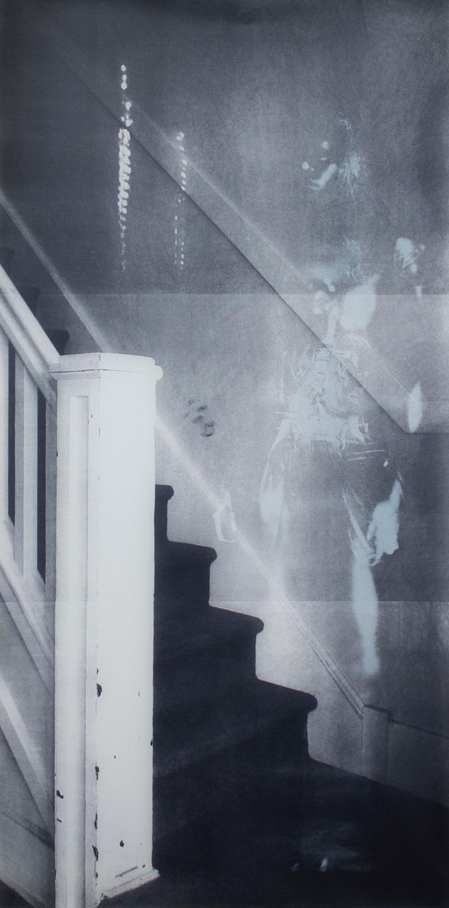
Darian Goldin Stahl, Stairway, Silkscreen, 84″×42″, 2014.
In the first complete exhibition that resulted from our collaboration, I used seven different photographic printmaking processes aimed at capturing the unseen: the quiet unease of daily life that results from the knowledge of the disease that grows inside.12 I have three main goals for this body of work. The first is to show the anxiety of the ill person by portraying a figure that is pulled apart—imagery which points to tension within the body. The second is to rehumanise the MRI scans by placing the figure within a context, such as his or her bedroom or hallway. The third and most important goal is to create work that is universal enough for the viewers to impart their own identity, while still being specifically about living daily with illness.
Scale also plays an important role within the work. Most of the pieces are printed to human scale so that the viewer can have a personal interaction with the body being portrayed. Entire figures are printed into domestic spaces that are life-sized, inviting the viewers to place themselves within the space as well. These pieces are presented unframed and set off of the wall—a gesture that breaks down the barrier between the art and the audience. Conversely, other pieces are printed very small or in book form, which forces the viewer to get up close or even handle the prints as a way of connecting with the artwork. Throughout the exhibition, measurement and scale symbols from the original digital set of MRI scans are a constant reminder of how the body can be quantified instead of qualified. I urge the viewer to consider the implications of measurements and data on the body, and broaden their point of view to see the figure as a whole agent (figure 2).

Darian Goldin Stahl, Doorway, Silkscreen, 84″×42″, 2013.
I have received feedback from the presentation of this body of work that points to the universality of the themes represented. Most of my time during an exhibition is spent with audience members who seek me out to tell me their personal story of illness. In particular, viewers who have an illness or have been scanned in a hospital are able to relate this work to their own experiences, and feel compelled to continue the narrative that DS and I have started by sharing their own. Other viewers have the experience of being a caregiver to an impaired family member, and find that our partnership mirrors their own efforts to empathise with and empower their loved one. Another group that I receive a lot of feedback from is the medical community. One neurologist confessed to me that she did not recognise the particular part of the brain I presented in an artwork, because it does not contain diagnosable areas for her specialty of MS. Because the work does not solely depict diagnosable images, but rather considers each scan as a part of a whole narrative, she was able to step back from her narrow scope of study and reconsider the entirety of the patient's body.
By joining the scans with the bodily impressions and placing them within a context, our traces combine through printmaking to construct a new figure, which is the amalgamation of our collaborative endeavours to give context to the medicalised body. I aim for viewers to identify with this figure and come to find that we all carry anxiety about the functionality of our bodies and, more broadly, our mortality. When audiences see my work, even though they do not know my sister, I hope they feel her and are filled with a shared, connecting reflection over the state of our ever-failing bodies.
How art reframes my experience—DS
DGS's art helps me to see my own MRIs as less alien. Of course, her art does not completely obliterate the schism that I feel between these visual representations of my interior and my own subjective experiences of illness, but it certainly helps to close the gap. Unlike the MRIs my physician chooses to interpret for me, DGS's work situates those MRIs within a representation of my body. By placing the MRIs of my interior within an exterior body, I can easily identify what part of my own body I am viewing when I see her art. Although this may seem like a minor point, there is a certain sort of embarrassment I feel whenever my physician shows me a representation of myself that I cannot identify as human much less my own brain or spine. MRIs are so abstract that they are nearly impossible to interpret by a layperson, much less identify with. DGS's art helps to put my body back together—interior with exterior and parts with the whole. Rather than hundreds of dissected pieces, her work shows an entire body stitched back together. Moreover, by making the art life-sized, I can physically stand next to it and see more precisely how the MRI maps onto my own body.
Of course, the body in DGS's art is not exactly mine. My body-in-art is but one more interpretation offered by an expert. Moreover, the body is not all mine, it is a hybrid body, composed of parts from both DGS and I. The surface impressions she creates fill out my exterior and yet are supposed to represent me. Although for some, this may take away from the authenticity of my bodily reconstruction, I see DGS's body as offering something important to my body's representation. By using her own body, DGS reminds me that my body and my identity do not exist in a vacuum. I am a person-in-relationship; my self-identity is constructed partially through my relationships and one of the most important relationships I have is with my sister. We share a physical resemblance as well as a common set of experiences. By merging her temporarily able-body with mine, I am also reminded that anyone could have MS. At present, few people can see that I have MS, because my symptoms are predominately invisible. Many people I have encountered are surprised to learn I have MS because they believed MS always resulted in obvious physical impairments. As I have come to learn, however, people with MS have a range of symptoms and some can be largely asymptomatic. DGS's art recalls that appearances can be deceiving.
Along with an exterior body, DGS's art also places my body into a particular configuration and within a physical space. Part of what makes the process of our collaboration therapeutic is that DGS bases her art off of my narrative accounts. Unlike my physician who rarely, if ever, asks about how I experience my illness or how it impacts my life, DGS takes my experiences and feelings seriously and uses them as the basis of her work. Each configuration of my body and each background space represent an experience that I have had and related to DGS. Knowing that my narrative will be expressed through fine art compels me to think more deeply about my own experiences as well as how I can make those experiences relatable to people who do not have MS. In short, DGS's art has made me into a storyteller. Over the years, I have become more comfortable telling others about my MS, because I have already told these same stories to DGS and seen them represented through her art. The potential for art and storytelling makes my frustrating experiences with MS and my physician slightly more tolerable, because they are a wellspring for education and creativity. Practice relating my experiences with others also empowers me to be more honest and direct with my clinical care providers. Whereas I am prone to being a passive recipient of medical care, my collaboration with DGS has empowered me to become more proactive in my interactions with physicians, so that I can be a collaborator in my own healthcare.
DGS's art has transformed how I view my MRI scans, and it has also altered how I feel undergoing those scans. Although I still dread the 3 h I spend restrained in that loud, narrow, claustrophobia-inducing tube, I also know the products of these scans are a new opportunity for creativity. I now anticipate my physician viewing these scans and updating me on my health, and later DGS and I viewing the scans and announcing which ones look frightening, ghostly, aethereal or comical and what each of those impressions reveals about my own personality or experiences. My MRIs are simultaneously a cold and objective tool used by my physicians to manage my health and as a source of subjective creativity and identity formation. These dual interpretations of my ill-body come close to how I relate to my own illness. At times, MS feels distant from me—a thing which has happened to me and over which I have little control. MS is an outside force, which can be managed by trained professionals and medication. Yet, at other times, I recognise that my MS is an indistinguishable part of me and who I have learned to be. To eradicate my MS would be to take something away from me—something which has connected me to my body and the bodies of others in a profound way.
Aims for the collaboration DS and DGS
As previously mentioned, the goals for this arts-based research are grounded in empathy creation. Although a specific person inspires these works, the figures in the prints are open enough for viewers to impart their own identity and experiences within the medical system. DGS's work speaks to the other side of the patient's life outside of the medical office. Unlike a doctor, DGS is not interested in pointing to diagnosable areas of the MRI scans. Instead, she uses the scans as a broader visual metaphor for living with disease. Although this artwork is specifically about DS and DGS's anticipation of disability, impending disability is a future most people will face if they live into old age. Knowing and understanding people with different bodies and abilities adds insight, dimension and value to the human condition.
Medical fine art may help people, particularly those with chronic illness, see the human body and illness differently. Artistic images often do what language cannot: they call us out of ourselves; they demand that we, the viewer, use our own interpretative skills. By sharing this art with the public, we hope to shape the way people view chronically ill bodies. By granting others the power of interpretation, we hope the images will help viewers to reconsider how they see illness and what it is like to live with disease. Depicting the worlds of bodily unease and anxiety together with the familiar world of everyday life enables the images to be more universally relatable. Likewise, we believe that patients who are given the opportunity to share in the power of interpretation will learn to see themselves differently, perhaps even multiply, and may begin to feel less objectified and alienated in the medical process.
Conclusion
Just as our bodies are multidimensional, their representations should be as well.
For obvious diagnostic purposes, clinical medicine favours a particular and singular interpretation of medical images. Medical imaging technologies are essential for clinical medicine; for the layperson, however, medical imaging technologies produce images of the body that are generally unrelatable and highly difficult to interpret. Moreover, such images may have the unintended side effect of alienating patients from their own bodies. By allowing for more anatomical interpreters, the body can take on new dimensions of meaning. Although in contemporary Western society, many tend to believe the hard science provide the truest or most accurate interpretation of the natural world, it cannot exhaust the meaning of the body. If we believe we are more than the sum of our parts, then we ought to allow for multiple and even varied interpretations of our bodies. Medical fine art is just one way we may express the inexhaustibility of the human body.
Footnotes
Competing interests None declared.
Provenance and peer review Not commissioned; externally peer reviewed.